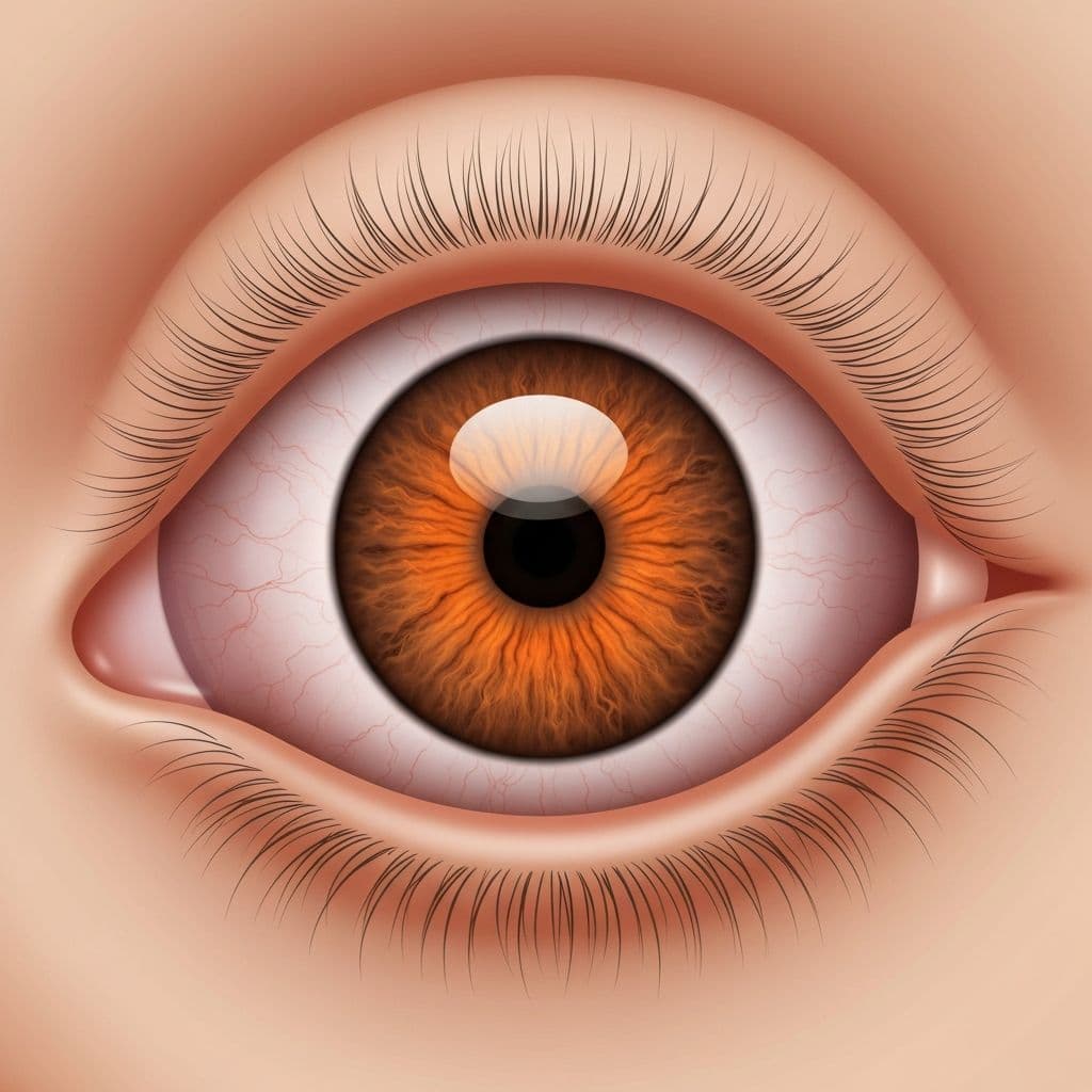
Recognize retinal detachment symptoms requiring emergency care. Learn about sudden vision changes, floaters, flashes, and when to seek immediate medical attention.
Retinal Detachment Symptoms: Essential Warning Signs and Emergency Response
Introduction
Retinal detachment is an ophthalmologic emergency requiring immediate medical attention. Retinal detachment symptoms, if recognized promptly and treated urgently, can often preserve vision, making knowledge of warning signs literally sight-saving. Understanding these symptoms and acting quickly can mean the difference between preserved vision and permanent blindness.
The retina, a light-sensitive tissue at the back of the eye, is essential for vision. When it separates from underlying blood vessels, nutrients and oxygen are cut off, causing rapid photoreceptor death. Without emergency intervention, complete vision loss in the affected eye typically results within days to weeks. This guide explores retinal detachment symptoms, risk factors, and critical emergency response protocols.
Table of Contents
- Retinal Anatomy and Function
- Early Warning Symptoms
- Progressive Symptoms
- Advanced Stage Symptoms
- Types of Retinal Detachment
- Risk Factors and High-Risk Groups
- Distinguishing Retinal Detachment from Other Conditions
- Emergency Response Protocols
- Frequently Asked Questions
- Conclusion
Retinal Anatomy and Function {#retinal-anatomy}
Understanding the Retina
The retina is a complex, multilayered tissue at the eye's back responsible for:
- Light detection: Photoreceptors (rods and cones) capture light
- Signal processing: Neural cells process visual information
- Signal transmission: Optic nerve carries information to brain
- Color and night vision: Cones provide color; rods provide low-light vision
Blood Supply Importance
The retina has two blood supply sources:
- Retinal blood vessels: Supply inner retinal layers
- Choroidal blood vessels: Supply outer photoreceptor layer
When detachment occurs, photoreceptors lose their blood supply, beginning to die within hours.
Why Detachment Is an Emergency
Time-critical process:
- Retinal cells begin dying within hours
- Death accelerates after 24-48 hours
- Complete vision loss can occur within 7-14 days if untreated
- Visual recovery depends on detachment duration before treatment
Early Warning Symptoms {#early-symptoms}
Floaters: The First Symptom
What are floaters?
Floaters are small specks or shadows drifting across visual field:
- Appearance: Thread-like strands, dots, or cobweb patterns
- Movement: Float with eye movement; slight lag when eyes stop
- Prevalence: Common, usually harmless
- Normal occurrence: Increase with age
Concerning floater characteristics:
- Sudden appearance: Multiple new floaters appearing suddenly
- Large numbers: Sudden increase in floater quantity
- Associated symptoms: Floaters plus other symptoms (see below)
- Accompanied by flashes: Especially concerning
Why floaters indicate problems:
When detachment begins, vitreous (gel inside eye) pulls on retina, causing:
- Vitreous hemorrhage (bleeding into vitreous)
- Blood cells appear as new floaters
- Sudden increase in floaters suggests serious pathology
Photopsia (Light Flashes)
Description:
Light flashes appear as lightning-like streaks at eye periphery:
- Location: Usually at eye edges, not central vision
- Trigger: Often occur with eye movement
- Duration: Brief, lasting seconds to minutes
- Frequency: May occur repeatedly
Mechanism:
Detaching retina tugs on vitreous, stimulating photoreceptors and creating false light perception.
Significance:
Flashes combined with floaters suggest retinal traction or early detachmentseek immediate care.
Importance of Prompt Recognition
Early intervention timing:
- Symptoms within hours warrant urgent evaluation
- Evaluation within 24 hours significantly improves outcomes
- Rhegmatogenous detachment requires same-day evaluation
- Treatment within days prevents vision loss
Progressive Symptoms {#progressive-symptoms}
Shadow or Curtain in Vision
Visual description:
- Shadow appearance: Dark area encroaching on visual field
- Location: Typically begins peripherally (edge of vision)
- Progression: Moves inward toward center
- Density: May appear as dim or dark shadow
- Progression rate: Can advance over hours to days
What causes the shadow:
As retina detaches, the separated portion no longer functions, creating a non-seeing area appearing as shadow or darkness.
Critical timing:
Shadow reaching the macula (central vision area) marks transition from preservable to potentially permanent vision loss.
Blurred or Distorted Vision
Symptoms:
- Vision becomes blurry or fuzzy
- Straight lines appear wavy (metamorphopsia)
- Images appear distorted
- Close visual tasks become difficult
- May affect one eye only
Cause:
Detachment affects focal point, refracting light incorrectly and distorting images.
Flashing Lights
Characteristics:
- More frequent, more intense than initial flashes
- May appear as lightning streaks
- Often accompanied by floater increase
- More noticeable in dim lighting
Significance:
Increasing flashes suggest progressive retinal tractiontime-critical symptom.
Advanced Stage Symptoms {#advanced-symptoms}
Significant Vision Loss
Extent of loss:
- Noticeable portion of visual field gone
- Large shadow occupying significant field area
- Central vision affected if macula involved
- Vision loss may progress over 24-48 hours
Experience:
- Like looking through partially blocked window
- Losing ability to perform normal tasks
- Difficulty reading, recognizing faces, driving
- Can progress to complete blindness in affected eye
Complete or Near-Complete Vision Loss
Final stage symptoms:
- Most or all vision lost in affected eye
- Only light perception may remain
- Occurs if macula is detached for extended period
- Largely irreversible if not surgically repaired
Why this occurs:
Photoreceptor death is progressive and eventually permanent after 7-14 days.
Types of Retinal Detachment {#types-detachment}
Rhegmatogenous Detachment (Most Common)
Characteristics:
- Cause: Break or tear in retina with fluid seeping behind
- Frequency: 90% of all detachments
- Symptoms: Typically sudden floaters, flashes, then shadow
- Risk: High-risk populations: high myopia, prior retinal surgery, family history
Tractional Detachment
Characteristics:
- Cause: Scar tissue pulls retina away
- Associated conditions: Diabetic retinopathy, severe eye trauma
- Symptom onset: Often gradual
- Vision impact: Variable depending on extent
Exudative Detachment
Characteristics:
- Cause: Fluid accumulates beneath retina without break
- Associated: Inflammation, infection, bleeding disorders
- Symptoms: Often less dramatic than rhegmatogenous
- Reversibility: May resolve with treatment of underlying cause
Risk Factors and High-Risk Groups {#risk-factors}
High Myopia (Nearsightedness)
Risk mechanism:
- Myopic eye has stretched, thinner retina
- Increased risk of retinal breaks
- Risk increases with higher myopia degrees
- Higher prevalence in Asian populations
Prior Eye Surgery
Surgical causes:
- Cataract surgery (slight increase)
- Refractive surgery (LASIK, PRK)
- Retinal surgery
- Glaucoma surgery
Family History
Genetic predisposition:
- Family history increases risk
- Suggests shared genetic factors
- May relate to myopia or retinal characteristics
Previous Retinal Detachment
Recurrence risk:
- 10-15% of patients experience recurrence
- May occur in same or other eye
- Usually requires additional treatment
- Vigilance for symptoms in both eyes important
Age and Other Factors
Additional risks:
- Age: Peak incidence 50-60 years
- Male gender: Higher incidence than females
- Trauma: Eye injury increases risk significantly
- Lattice degeneration: Thin retinal areas predispose
- Posterior vitreous detachment: Natural separation of vitreous from retina increases risk
Distinguishing Retinal Detachment from Other Conditions {#differentiation}
Retinal Detachment vs. Posterior Vitreous Detachment
Posterior Vitreous Detachment (PVD):
- Common: Happens to most people eventually
- Cause: Natural aging process separates vitreous from retina
- Symptoms: Floaters and flashes possible
- Risk: Can lead to retinal detachment if tear occurs
- Urgency: Requires evaluation but often benign
Distinguishing factors:
- Detachment has progressive vision loss; PVD doesn't
- Detachment may have Weiss ring (ring floater specific to PVD)
- Detachment affects visual field; PVD doesn't
Retinal Detachment vs. Retinal Tear
Retinal Tear:
- Definition: Rip in retina without detachment
- Progression: May progress to detachment (20-30% risk)
- Treatment: Preventive laser or cryotherapy recommended
- Urgency: Same-day evaluation necessary
Retinal Detachment vs. Stroke
Amaurosis Fugax (Transient Vision Loss):
- Cause: Temporary blood vessel blockage
- Duration: Complete recovery of vision within hours
- Symptom: Unlike detachment's progressive loss
- Risk: Indicates cardiovascular disease risk
Retinal Detachment vs. Migraine Aura
Migraine visual symptoms:
- Duration: Temporary, typically 20-30 minutes
- Pattern: Often zigzag patterns
- Other symptoms: Usually followed by headache
- Recovery: Complete visual recovery
Detachment differs:
- Progressive rather than temporary
- Shadow rather than scintillating patterns
- No headache component
- No visual recovery without treatment
Emergency Response Protocols {#emergency-response}
Immediate Actions
When symptoms occur:
- Stop what you're doing: Stop driving, cease activities
- Seek emergency care immediately: Don't wait for appointment
- Go to emergency room or ophthalmology urgent care: Not regular urgent care
- Describe symptoms clearly: Emphasize sudden floaters, flashes, shadow
- Don't apply pressure: Don't press on eye
- Avoid straining: Don't lift heavy objects or strain
How to Access Emergency Ophthalmology
Options:
- Hospital emergency room: Can access ophthalmology consultation
- Urgent care ophthalmology: If available in your area
- On-call ophthalmologist: Through your primary eye doctor
- Retinal specialty center: Often available for emergencies
Diagnostic Procedures
During emergency evaluation:
- Visual acuity testing: Checks vision level
- Slit lamp examination: Magnified eye inspection
- Dilated retinal exam: Essential for diagnosis
- Ultrasound (B-scan): If view obscured by blood or opacity
- OCT imaging: High-resolution retinal imaging if available
Frequently Asked Questions {#faqs}
Can retinal detachment cause permanent blindness?
Yes, if untreated:
- Untreated detachment leads to complete vision loss
- Surgery success depends on treatment timing
- Macula-on detachment (untouched macula): 80-90% excellent vision recovery
- Macula-off detachment: 50-60% retain useful vision even with surgery
How quickly does retinal detachment progress?
Variable timing:
- Initial symptoms: Floaters and flashes (immediate)
- Shadow development: Hours to days
- Macula involvement: Often within days if untreated
- Complete loss: 7-14 days if untreated
- Rapid progression requires emergency care
Are all floaters concerning?
No:
- Age-related floaters are common and benign
- Sudden increase in quantity warrants evaluation
- Floaters with flashes are more concerning
- Any sudden vision change requires evaluation
What is the success rate of retinal detachment surgery?
Outcomes:
- Primary repair success: 80-90% with modern techniques
- Vision recovery: Depends on macula status and duration
- Some patients require multiple procedures
- Early detection dramatically improves outcomes
Can retinal detachment happen in both eyes?
Risk of bilateral detachment:
- Uncommon but possible
- Higher risk if high myopia or familial history
- Second eye monitoring important
- Preventive treatments available for high-risk patients
Conclusion
Retinal detachment symptomssudden floaters, photopsia, shadow in visionrepresent an ophthalmologic emergency requiring immediate intervention. The critical importance of recognizing these warning signs cannot be overstated: treatment within hours dramatically improves outcomes, while delays measured in days result in permanent vision loss.
If you experience sudden floaters, flashing lights, or a shadow encroaching on your visual field, seek emergency ophthalmologic care immediately. Your vision may depend on it. Modern surgical techniques offer excellent outcomes when treatment is prompt, making early recognition and rapid response your most powerful tools for preserving sight.